
More Information
Submitted: 14 February 2020 | Approved: 17 February 2020 | Published: 18 February 2020
How to cite this article: Touimi SH, Daoudi S, Mbarki I, Adrif I, Elkacemi H, et al. An unusual case of a maxillary sinonasal neuroendocrine carcinoma. J Clin Med Exp Images. 2020; 4: 001-001.
DOI: 10.29328/journal.jcmei.1001014
Copyright License: © 2020 Pérez J, et al. This is an open access article distributed under the Creative Commons Attribution License, which permits unrestricted use, distribution, and reproduction in any medium, provided the original work is properly cited.

An unusual case of a maxillary sinonasal neuroendocrine carcinoma
Touimi SH1*, Daoudi S2, Mbarki I1, Adrif I2, Elkacemi H1, Elmajjaoui S1, Kebdani T1, Errihani H2 and Benjaafar N1
1Department of Radiotherapy, National Institute of Oncology, Rabat, Morocco
2Department of Oncology, National Institute of Oncology, Rabat, Morocco
*Address for Correspondence: Touimi SH, Department of Radiotherapy, National Institute of Oncology, Rabat, Morocco, Email: [email protected]
It’s a 24 years old female patient who presented with rhinological burning pain evolving since 1 year. She didn’t consult until a blistering lesion filled half of the oral cavity. The initial biopsy of the tumor was interpreted as a round cell tumor process.
It’s a 24 years old female patient who presented with rhinological burning pain evolving since 1 year. She didn’t consult until a blistering lesion filled half of the oral cavity. The initial biopsy of the tumor was interpreted as a round cell tumor process. An immuno-histochemical complement showed a poorly differenciated neuroendocrine carcinoma, (Pancytokeratine+, Ki67 at 30%, Chromogranine+, Synaptophysine-).
CT scan showed a localy advanced maxilary nasal sinus tumorof 74mm in the greatest diameter (Figure 1). The remainder of the staging didn’t reveal any metastases. The patient received 3 courses of chemotherapy with Etoposod-cisplatin. The clinical and radiological evaluations showed a progression of disease
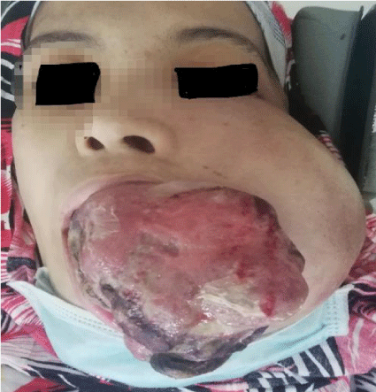
Figure 1: A 24 years old patient with a maxillary sinonasal carcinoma.