
More Information
Submitted: August 14, 2022 | Approved: August 29, 2022 | Published: August 30, 2022
How to cite this article: Mouftah B, Amine S, Jaafar F, Ayoub M, Tarik K, et al. Huge median prostatic lobe: a interesting case of BPH. J Clin Med Exp Images. 2022; 6: 006-006.
DOI: 10.29328/journal.jcmei.1001026
Copyright License: © 2022 Mouftah B, et al. This is an open access article distributed under the Creative Commons Attribution License, which permits unrestricted use, distribution, and reproduction in any medium, provided the original work is properly cited.

Huge median prostatic lobe: a interesting case of BPH
Babty Mouftah*, Slaoui Amine, Fouimtizi Jaafar, Mamad Ayoub, Karmouni Tarik, El Khadder Khalid, Koutani Abdellatif and Ibn Attya Ahmed
Department of Urology B, IBN SINA Hospital, Mohammed V University, Rabat, Morocco
*Address for Correspondence: Babty Mouftah, Department of Urology B, IBN SINA Hospital, Mohammed V University-Rabat, Morocco, Email: [email protected]
Benign Prostatic Hyperplasia (BPH) refers to the nonmalignant growth or hyperplasia of prostate tissue and is a common cause of lower urinary tract symptoms in men [1]. Its management is essentially medical. Surgery is performed after failure of medical treatment or in case of complicated BPH. We report the case of a huge median lobe occupying almost the entire bladder lumen. This is the case of a 79 - year-old patient, a non-smoker, who had a history of terminal macroscopic hematuria. The patient presented with lower urinary tract symptoms in the last 2 years, treated first by medicine with poor response. On digital rectal examination, the enlarged prostate was homogeneous and regular, exceeding 60 g. The biological workup was unremarkable except for a total PSA level of 16 ng/ml.
A first abdominopelvic CT scan objectified a vesico-prostatic mass, hence the realization of a diagnostic cystoscopy.
The resectoscope that we had at our disposal could not penetrate the bladder despite a 50 g resection of the median lobe.
We decided to do a second abdominopelvic CT scan, which showed a voluminous prostate estimated at 363 g with a huge median lobe. Transversal prostatic adenomectomy is indicated Figure 1.
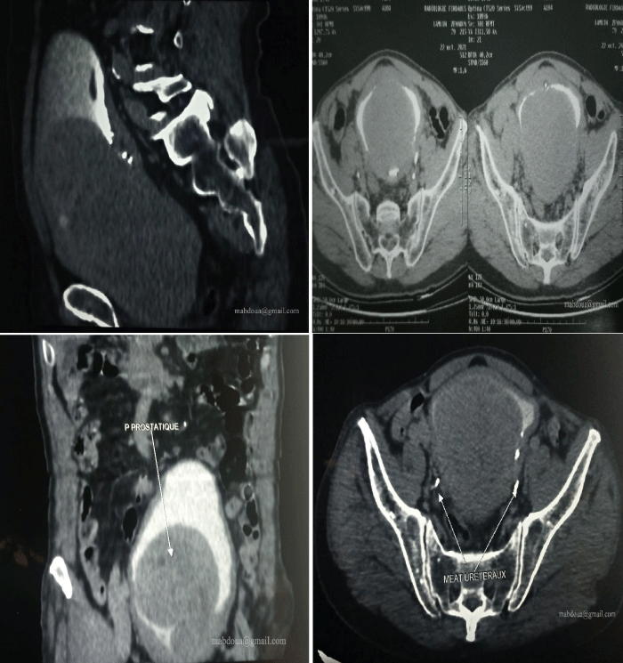
Figure 1: Different sections showing a median lobe occupying almost the entire bladder lumen.