
More Information
Submitted: March 08, 2023 | Approved: March 16, 2023 | Published: March 17, 2023
How to cite this article: Bajaj A. Pomaded and unctuous- spindle cell lipoma. J Clin Med Exp Images. 2023; 7: 001-003.
DOI: 10.29328/journal.jcmei.1001027
Copyright License: © 2023 Bajaj A. This is an open access article distributed under the Creative Commons Attribution License, which permits unrestricted use, distribution, and reproduction in any medium, provided the original work is properly cited.

Pomaded and unctuous- spindle cell lipoma
Anubha Bajaj*
Histopathologist, A.B. Diagnostics, New Delhi, India
*Address for Correspondence: Anubha Bajaj, Histopathologist, A.B. Diagnostics, New Delhi, India, Email: [email protected]
Spindle cell lipoma and pleomorphic lipoma emerge as benign, adipocytic neoplasms representing a morphologic continuum of a singular neoplasm. In addition to myofibroblastoma and cellular angiofibroma, spindle cell lipoma or pleomorphic lipoma configures as a constituent of chromosome 13q/RB1 family of tumors. Initially scripted by Enzinger and Harvey in 1975 with an obsolete terminology of dendritic fibromyxolipoma, aforesaid adipocytic neoplasms articulate an estimated 1.5% of lipomatous tumors. In contrast to conventional lipoma, spindle cell lipoma or pleomorphic lipoma is an uncommonly discerned variant, demonstrating a proportion of 1: 60.
Spindle cell lipoma is constituted of varying quantities of mature adipocytes, bland spindle-shaped cells, and ropy collagen fibers. In addition to aforesaid constituents, pleomorphic lipoma exemplifies pleomorphic and multinucleated floret-like giant cells.
Spindle cell lipoma or pleomorphic lipoma commonly incriminates middle-aged to elderly subjects between 45 years to 70 years. Tumefaction is exceptional < 20 years. A male predominance is observed with < 10% of tumors occurring in females [1,2]. Several neoplasms depict monosomy along with partial loss of chromosome 13 and chromosome 16. Loss of RB1 genetic locus may be encountered [1,2].
Spindle cell lipoma or pleomorphic lipoma emerges as a superficial lesion. Intramuscular and dermal neoplasms appear infrequently. Deep-seated, soft-tissue lesions are extremely exceptional [2,3]. The majority (~80%) of tumefaction is confined to the subcutaneous tissue of the posterior cervical region, dorsum, or shoulders. Around 20% of lesions occur within diverse sites such as the face, oral cavity, trunk, or extremities. Generally, incriminated females depict lesions confined to atypical locations [2,3]. Characteristically, spindle cell lipoma or pleomorphic lipoma is associated with partial or comprehensive deletion of chromosome 13 or chromosome 16. Besides, breakpoint clusters surrounding chromosome 13q14 may ensue, a location where the RB1 gene is situated.
Familial instances demonstrating multiple lesions of spindle cell lipoma or pleomorphic lipoma are documented [2,3].
Generally solitary, miniature, and asymptomatic, spindle cell lipoma or pleomorphic lipoma appears as a mobile, subcutaneous lesion of significant duration. The Tumour magnitude is < 5 cm. Tumefaction is commonly confined to the shoulder, posterior cervical, or upper dorsal region [2,3].
Upon gross examination, a well-circumscribed, elliptical tumefaction appears to be confined within the subcutaneous tissue. The Cut surface is yellow to tan with foci of grey/white or myxoid tumor zones. In contrast to conventional lipoma, the texture is firm. Dermal and intramuscular tumors may exhibit an infiltrative tumor countenance.
Frozen section examination exhibits aggregates of mature adipocytes intermixed with bland spindle-shaped cells [3,4].
Upon cytological examination, an admixture of mature adipocytes, uniform spindle-shaped cells, and collagen fibers are encountered. Spindle-shaped cells are imbued with fusiform to ovoid nuclei and demonstrate inadequately defined bipolar cytoplasmic processes. Nuclear grooves may be discerned. Characteristically, nuclear pleomorphism or mitotic figures are absent. Pleomorphic lipoma may delineate multinucleated giant cells. Accompanying mast cells are commonly disseminated within a myxoid background [3,4].
Upon microscopy, the tumor is composed of an admixture of mature adipocytes, bland spindle-shaped cells, and hyalinised, rope-like collagen fibers. Pleomorphic lipoma depicts dispersed multinucleated stromal giant cells and floret-like cells. Mast cells are frequently disseminated within the myxoid substance. Spindle-shaped cells may appear as short, stubby, or blunt and occasionally delineate palisading. Lipoblasts are exceptional. Generally, features such as infiltrative tumor growth, focal necrosis, atypical spindle-shaped cells, pleomorphic lipoblasts, and significant mitotic activity are absent [3,4].
Spindle cell lipoma or pleomorphic lipoma demonstrate cogent subtypes as ~‘fat poor’ or ‘fat-free’ wherein the variant is constituted of minimal (< 5%) to absent mature adipose tissue cells. ~myxoid subtype is comprised of the abundant myxoid stroma. ~pseudo-angiomatous subtype demonstrates distended vascular channels layered with endothelial cells admixed with floating tumor nodules. ~a plexiform variant is documented [3,4]. Exceptionally, focal cartilaginous or osseous metaplasia may ensue. Besides, foci of extramedullary hematopoiesis may be discerned. Upon ultrastructural examination, spindle cell lipoma is constituted of spindle-shaped cells exhibiting features of fibroblasts. Besides, non-membrane bound lipid droplets are discerned, indicative of neoplastic elements of the pre-lipoblast stage [3,4] Figures 1-3.

Figure 1: Spindle cell lipoma demonstrating aggregates of bland spindle-shaped cells admixed with ropy collagen and mature adipose tissue cells [6].
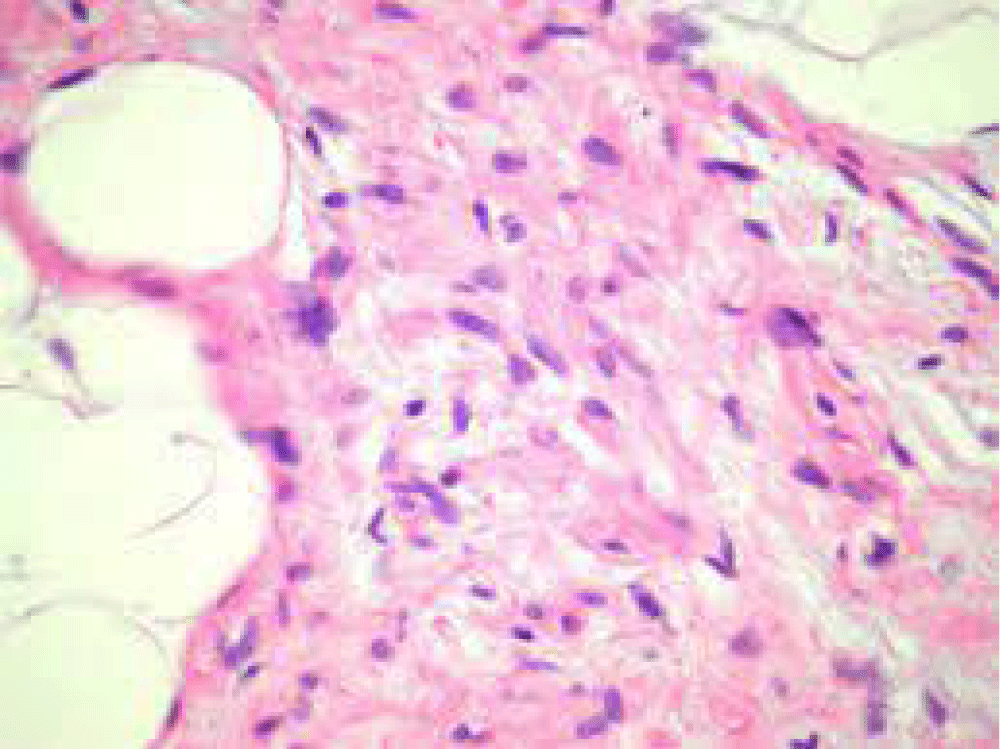
Figure 2: Spindle cell lipoma exhibiting bundles of uniform spindle-shaped cells intermingled with ropy collagen and mature adipose tissue cells [7].
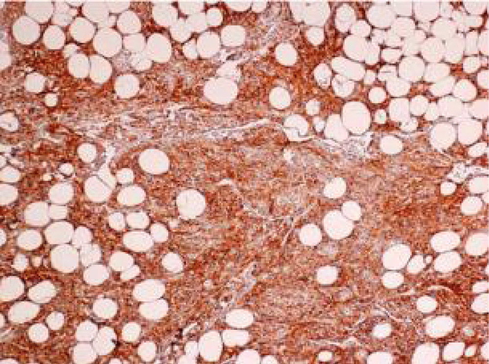
Figure 3: Spindle cell lipoma immune reactive to CD10 [8].
Spindle cell lipoma or pleomorphic lipoma is immune reactive to CD34 and vimentin. Tumour depicts the loss of expression of nuclear RB1 protein. Spindle cell lipoma or pleomorphic lipoma is immune and non-reactive to keratin, S100 protein, SOX10, smooth muscle actin (SMA), desmin, STAT6, or estrogen receptor [4,5].
Spindle cell lipoma and pleomorphic lipoma require segregation from neoplasms such as fibro lipoma, pleomorphic liposarcoma, myxoid liposarcoma, low-grade myxofibrosarcoma, a lipomatous variant of the solitary fibrous tumor, mammary type myofibroblastoma, cellular angiofibroma, dermatofibrosarcoma protuberans, schwannoma or neurofibroma. Spindle cell lipoma can be appropriately discerned as a superficial, well-circumscribed, adipocytic neoplasm. Lesions are characteristically exemplified upon the shoulder, posterior cervical region, or dorsal area. Middle-aged to elderly, male subjects are commonly incriminated. Neoplasm is immune-reactive to CD34. Tumour cells depict the loss of the RB1 gene [4,5]. Magnetic resonance imaging (MRI) demonstrates nonspecific features and manifests a variable appearance wherein neoplasm may occur as a completely non-lipogenic tumor to a conventional lipoma-like lesion. The investigative modality is beneficial in determining the extent of the lesion. Additionally, the ‘fat-free’ variant may be misinterpreted as a sarcoma. Spindle cell lipoma or pleomorphic lipoma can be appropriately managed with conservative manoeuvers as singular surgical eradication of neoplasm [4,5]. Spindle cell lipoma or pleomorphic lipoma emerges as a benign lesion. Possible localized or distant metastasis is absent. Besides, localized tumor reoccurrence is exceptional despite incomplete surgical resection of neoplasm [4,5].
- Tamamine S, Himejima T, Mitsui T, Masuoka H, Hihara M, Kakudo N. A case of occipital spindle cell lipoma: a case report. J Surg Case Rep. 2022 Nov 26;2022(11):rjac544. doi: 10.1093/jscr/rjac544. PMID: 36452288; PMCID: PMC9701562.
- Ben Salah N, Lahouel I, Manaa L, Youssef M, Belhadjali H, Zili J. Spindle cell lipoma: An uncommon variant of lipoma affecting the foot sole. Clin Case Rep. 2022 Feb 13;10(2):e05455. doi: 10.1002/ccr3.5455. PMID: 35198205; PMCID: PMC8841022.
- AlRashed R, Albdah A, Alsannaa F. A case report of spindle cell lipoma. Ann Med Surg (Lond). 2022 Jun 13;79:103960. doi: 10.1016/j.amsu.2022.103960. PMID: 35860096; PMCID: PMC9289322.
- Biagiotti J, Khan K, Ayrapetyan M, Wood J, Bhatt N. Fat-Free Spindle Cell Lipoma of the Scalp: Lipoma Without a Lipogenic Component. J Craniofac Surg. 2022 Jun 1;33(4):e429-e431. doi: 10.1097/SCS.0000000000008359. Epub 2021 Nov 5. PMID: 34743158.
- Engwall-Gill AJ, Xiong M, Bray SMC. Comprehensive Review of Reported Nonclassical Spindle Cell Lipoma Presentations and a Unique Case Report. Plast Reconstr Surg Glob Open. 2022 Aug 18;10(8):e4462. doi: 10.1097/GOX.0000000000004462. PMID: 35999878; PMCID: PMC9390821.
- Image I Courtesy: mypathologyreport.ca
- Image 2 Courtesy: Dermnet NZ
- Image 3 Courtesy: Research gate.