
More Information
Submitted: May 16, 2022 | Approved: August 01, 2022 | Published: August 02, 2022
How to cite this article: Daly H, Bouallegue A, Horchani A. A rare variant of the radial artery was discovered during a pseudoaneurysm of the brachial artery in a hemodialysis patient. J Clin Med Exp Images. 2022; 6: 003-005.
DOI: 10.29328/journal.jcmei.1001025
Copyright License: © 2022 Daly H, et al. This is an open access article distributed under the Creative Commons Attribution License, which permits unrestricted use, distribution, and reproduction in any medium, provided the original work is properly cited.

A rare variant of the radial artery was discovered during a pseudoaneurysm of the brachial artery in a hemodialysis patient
Hafedh Daly1*, Abdelbaki Bouallegue2 and Amira Horchani3
1Cardiovascular Surgery Department, Regional Hospital of Gafsa, Tunisia
2Nephrology and Hemodialysis Department, Regional Hospital of Gafsa, Tunisia
3Faculty of Pharmacy, Monastir, Tunisia
*Address for Correspondence: Hafedh Daly, Cardiovascular Surgery Department, Regional Hospital of Gafsa, Tunisia, Email: [email protected]
The radial artery shows great anatomical variability with respect to its origin [1]. Generally, its origin is located in the cubital fossa at the level of the neck of the radius [2]. However, the artery may have a high origin from the brachial artery or even the axillary artery [1].
We report a case with the presence of two radial arteries, one which arises from the axillary artery and one which normally arises from the brachial artery.
This is a 59 - year-old patient, on chronic hemodialysis for 7 months from a left radial fistula, who presented to the emergency room for the appearance of a throbbing and painful mass in the left arm, evolving for 3 days, without the notion of trauma. Doppler ultrasound of the upper limb showed the presence of a pseudoaneurysm of the left brachial artery. The CT angiography confirmed both the diagnosis of brachial pseudoaneurysm and the presence of two radial arteries; one of high birth from the axillary artery and one of usual birth from the brachial artery (Figures 1-4). The patient underwent emergency surgery by approaching the brachial artery at the level of the arm, surgical exploration showed the presence of a giant false aneurysm (Figure 5) and a 2 mm breach of the brachial artery which was sutured (Figure 6). The postoperative course was simple.
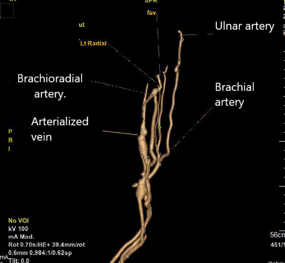
Figure 1: The CT angiography of the left upper limb shows the high birth of the left radial artery (brachioradial artery), the brachial artery gives 3 branches: the ulnar artery, the interosseous artery, and a second radial artery which is small.
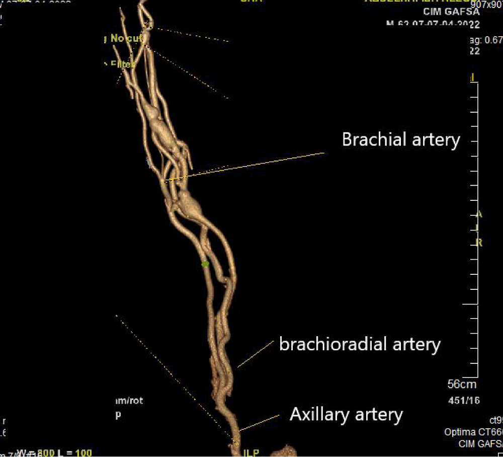
Figure 2: The anatomopathological examination concluded to cortico-adrenal tissue at the level of the spermatic cord with hypo spermatogenesis at the level of testicular sampling.
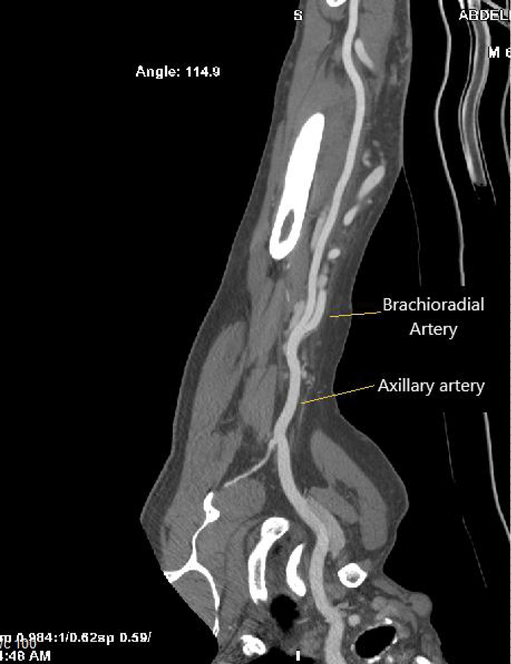
Figure 3: The CT angiography of the left upper limb which shows the upper birth of the left radial artery (brachioradial artery) from the axillary artery and the pseudoaneurysm of the brachial artery.
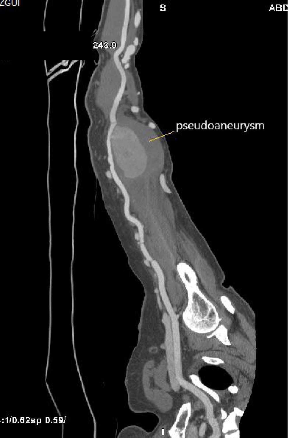
Figure 4: The CT angiography of the left upper limb which shows the upper birth of the left radial artery (brachioradial artery) from the axillary artery and the pseudoaneurysm of the brachial artery.
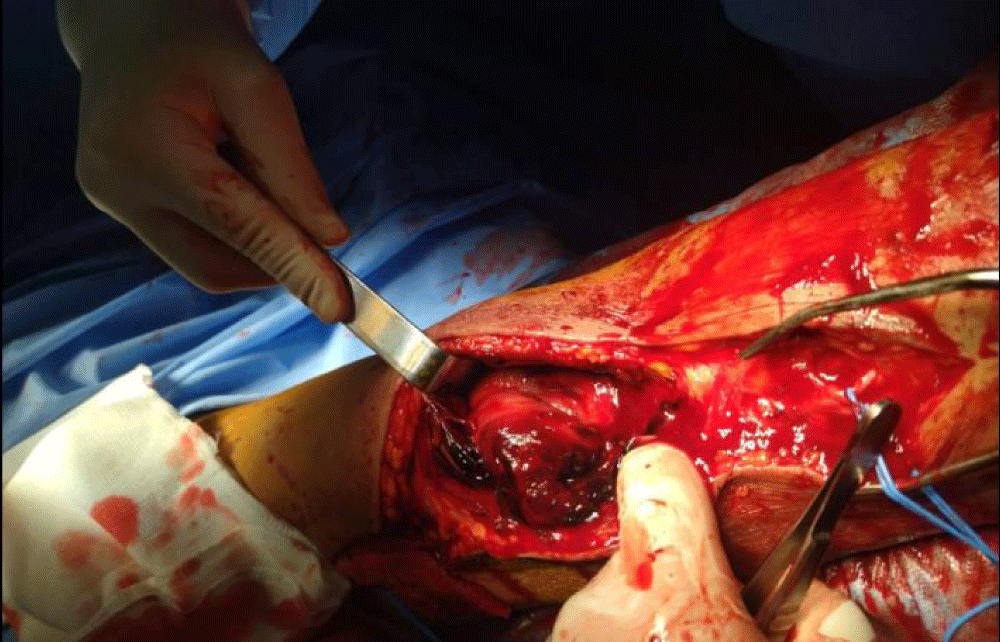
Figure 5: Intraoperative view showing the pseudoaneurysm.
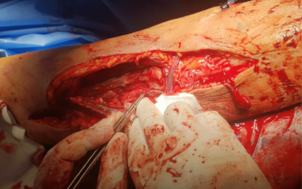
Figure 6: Intraoperative view showing suturing of the brachial artery breach.
Anatomical variations of the radial artery are relatively rare, with an incidence of around 0.2% [3]. These variations are represented by: a radial artery that detaches 76 mm below the intercondylar line of the humerus and which emerges under the tendon of the pronator teres muscle to end in the deep palmar arch [3], a branch unusual superficial radial artery that detaches before entering the palm passing between the two heads of the first dorsal interosseus to finally located on the dorsum of the hand [4], a rare and unique abnormal medial branch of the spiral radial artery around the flexor carpi radialis [5], compression of the median nerve at the carpal tunnel by a superficial and aberrant branch of the radial artery [6], the ulnar origin of the radial artery, split radial artery and finally, by the passage of a radial artery under the pronator teres muscle [7].
The arteries for the upper limbs derive mainly from the seventh cervical intersegmental artery. These arteries initially distribute to each limb bud by uniting with an axial artery which develops along the central axis of the bud. The axial artery gives off a brachial artery for the arm and an anterior interosseous artery for the forearm. The other arteries of the upper limb, including the radial and ulnar arteries, develop as sprouting from the axial artery [8].
At the first stage of the formation of the upper limb, the dominant vessel is the subclavian artery which continues thereafter with the axillary artery, the brachial artery, and the interosseous artery. It is only at a later stage of development that the ulnar and radial arteries appear [9,10].
Congenital vascular malformations can occur at many stages during the process of embryonic development. These malformations most often result from an absence of regression of certain primitive elements, or alternatively from an inappropriate regression of an element [8].
An aberrant birth of the radial artery is relatively rare, its discovery is often fortuitous, during its removal for coronary artery bypass or during vascular imaging.
- Adachi B. Das Arteriensystem der Japaner. 1928; 285-356. Maruzen, Kyoto 1.
- Standring S. Gray’s Anatomy: The Anatomical Basis of Clinical Practice. 40e. Édimbourg, Londres: Churchill Livingstone; Grays Anatomy: The Anatomical Basis of Clinical Practice. 2008; 905-906.
- Wysiadecki G, Polguj M, Haładaj R, Topol M. Low origin of the radial artery: a case study including a review of literature and proposal of an embryological explanation. Anat Sci Int. 2017 Mar;92(2):293-298. doi: 10.1007/s12565-016-0371-9. Epub 2016 Sep 15. PMID: 27631096; PMCID: PMC5315727.
- Nayak SB, Kumar N, Aithal Padur A, Shetty SD. Variant Superficial Branch of Radial Artery along with Supplementary Tendons on the Dorsum of the Hand and Their Surgical Implications. Case Rep Surg. 2016;2016:9581759. doi: 10.1155/2016/9581759. Epub 2016 Oct 5. PMID: 27818829; PMCID: PMC5069369.
- Wadhwa S, Tomar V. Anomalous Medial Branch of Radial Artery: A Rare Variant. Acta Medica (Hradec Kralove). 2016;59(3):100-103. doi: 10.14712/18059694.2016.98. PMID: 27770839.
- Kokkalis ZT, Tolis KE, Megaloikonomos PD, Panagopoulos GN, Igoumenou VG, Mavrogenis AF. Aberrant Radial Artery Causing Carpal Tunnel Syndrome. Arch Bone Jt Surg. 2016 Jun;4(3):282-4. PMID: 27517078; PMCID: PMC4969379.
- Yatzouta C. Bulletins and Memoirs of the Paris Anthropology Society, VII Series. 1927; 8: 31-38.
- Larsen W. Embryologie humaine. 2017; 325.
- Singer E. Embryological pattern persisting in the arteries of thearm Anat Rec. 1933; 55: 403-409.
- Standring S. Gray's anatomy: the anatomical basis of clinical practice Edinburgh: Churchill Livingstone. 2008; 905-906.