Figure 1
Ketamine-related uropathy and cholangiopathy
Sung-Yuan Hu*, Yi-Hsuan Chen and Shian-Shiang Wang
Published: 18 March, 2021 | Volume 5 - Issue 1 | Pages: 002-002

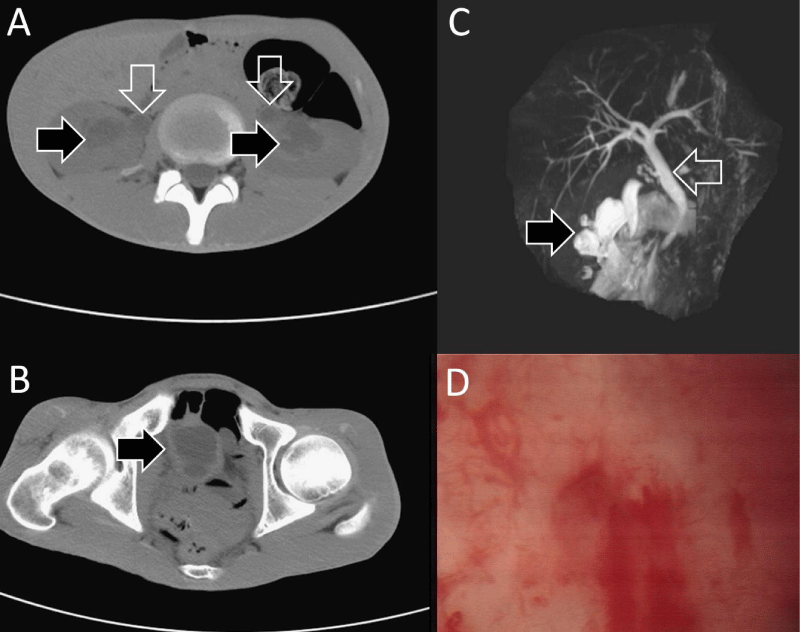
Figure 1:
Bilateral hydronephrosis (black arrows in panel A), hydroureter (white arrows in panel A), irregular thickened wall of urinary bladder (black arrow in panel B), fusiform common bile duct with distal stenosis (panel C), and ulcerative cystitis (panel D).
Read Full Article HTML DOI: 10.29328/journal.jcmei.1001018 Cite this Article Read Full Article PDF
More Images
Similar Articles
-
Cystic Micronodular Thymoma. Report of a CaseMona Mlika*,Adel Marghli ,Faouzi Mezni. Cystic Micronodular Thymoma. Report of a Case . . 2017 doi: 10.29328/journal.jcmei.1001001; 1: 001-004
-
Pulmonary Infarction Mimicking An Aspergilloma In A Heart Transplant RecipientAntonacci F*,Belliato M,Bortolotto C,Di Perna D,Dore R,Orlandoni G,D’Armini AM. Pulmonary Infarction Mimicking An Aspergilloma In A Heart Transplant Recipient . . 2017 doi: 10.29328/journal.jcmei.1001002; 1: 005-006
-
Recurrent Peripheral Ameloblastoma of the Mandible: A Case ReportAngela Jordão Camargo*,Mayara Cheade,Celso Martinelli,Plauto Christopher Aranha Watanabe. Recurrent Peripheral Ameloblastoma of the Mandible: A Case Report. . 2017 doi: 10.29328/journal.jcmei.1001003; 1: 007-010
-
Tumours of the Uterine Corpus: A Histopathological and Prognostic Evaluation Preliminary of 429 PatientsJorge F Cameselle-Teijeiro*,Javier Valdés-Pons,Lucía Cameselle-Cortizo,Isaura Fernández-Pérez,MaríaJosé Lamas-González,Sabela Iglesias-Faustino,Elena Figueiredo Alonso,María-Emilia Cortizo-Torres,María-Concepción Agras-Suárez,Araceli Iglesias-Salgado,Marta Salgado-Costas,Susana Friande-Pereira,Fernando C Schmitt. Tumours of the Uterine Corpus: A Histopathological and Prognostic Evaluation Preliminary of 429 Patients . . 2017 doi: 10.29328/journal.jcmei.1001004; 1: 011-019
-
The Risk Factors for Ankle Sprain in Cadets at a Male Military School in Iran: A Retrospective Case-control StudyFarzad Najafipour*,Farshad Najafipour,Mohammad Hassan Majlesi,Milad Darejeh. The Risk Factors for Ankle Sprain in Cadets at a Male Military School in Iran: A Retrospective Case-control Study. . 2017 doi: 10.29328/journal.jcmei.1001005; 1: 020-026
-
Magnetic Resonance Imaging Can Detect Symptomatic Patients with Facet Joint Pain. A Retrospective AnalysisWolfgang Freund*,Frank Weber,Reinhard Meier,Stephan Klessinger. Magnetic Resonance Imaging Can Detect Symptomatic Patients with Facet Joint Pain. A Retrospective Analysis. . 2017 doi: 10.29328/journal.jcmei.1001006; 1: 027-036
-
Secondary Onychomycosis Development after Cosmetic Procedure-Case ReportMariusz Dyląg*,Emilia Flisowska,Patryk Bielecki,Maria Kozioł-Gałczyńska,Weronika Jasińska. Secondary Onychomycosis Development after Cosmetic Procedure-Case Report . . 2017 doi: 10.29328/journal.jcmei.1001007; 1: 037-045
-
Andy Gump deformityPirabu Sakthivel*,Chirom Amit Singh,Chandra Sharma. Andy Gump deformity. . 2017 doi: 10.29328/journal.jcmei.1001008; 1: 046-047
-
The Death of a Baby from the Congenital Anomalies of the Urinary TractAstrit Gashi M*,Gent Sopa,Ilir Kadiri,Majlinda Bala,Petrit Pupa. The Death of a Baby from the Congenital Anomalies of the Urinary Tract. . 2018 doi: 10.29328/journal.jcmei.1001009; 2: 001-002
-
New technique of imaging cellular change to squmous cells metaplsia of cervixSalwa Samir Anter*. New technique of imaging cellular change to squmous cells metaplsia of cervix . . 2019 doi: 10.29328/journal.jcmei.1001010; 3: 001-001
Recently Viewed
-
Effects of dietary supplementation on progression to type 2 diabetes in subjects with prediabetes: a single center randomized double-blind placebo-controlled trialSathit Niramitmahapanya*,Preeyapat Chattieng,Tiersidh Nasomphan,Korbtham Sathirakul. Effects of dietary supplementation on progression to type 2 diabetes in subjects with prediabetes: a single center randomized double-blind placebo-controlled trial. Ann Clin Endocrinol Metabol. 2023: doi: 10.29328/journal.acem.1001026; 7: 00-007
-
Physical Performance in the Overweight/Obesity Children Evaluation and RehabilitationCristina Popescu, Mircea-Sebastian Șerbănescu, Gigi Calin*, Magdalena Rodica Trăistaru. Physical Performance in the Overweight/Obesity Children Evaluation and Rehabilitation. Ann Clin Endocrinol Metabol. 2024: doi: 10.29328/journal.acem.1001030; 8: 004-012
-
Hypercalcaemic Crisis Associated with Hyperthyroidism: A Rare and Challenging PresentationKarthik Baburaj*, Priya Thottiyil Nair, Abeed Hussain, Vimal MV. Hypercalcaemic Crisis Associated with Hyperthyroidism: A Rare and Challenging Presentation. Ann Clin Endocrinol Metabol. 2024: doi: 10.29328/journal.acem.1001029; 8: 001-003
-
The effect of frequency of sexual intercourse on coronary artery diseaseMehdi Karasu*,Özkan Karaca,Mehmet Ali Kobat,Tarık Kıvrak,Mehmet İkbal İpek. The effect of frequency of sexual intercourse on coronary artery disease. Arch Vas Med. 2022: doi: 10.29328/journal.avm.1001015; 6: 001-004
-
A Case Report on Paradoxical EmboliYou Li* and Jason Wheeler. A Case Report on Paradoxical Emboli. Arch Vas Med. 2024: doi: 10.29328/journal.avm.1001019; 8: 004-007
Most Viewed
-
Impact of Latex Sensitization on Asthma and Rhinitis Progression: A Study at Abidjan-Cocody University Hospital - Côte d’Ivoire (Progression of Asthma and Rhinitis related to Latex Sensitization)Dasse Sery Romuald*, KL Siransy, N Koffi, RO Yeboah, EK Nguessan, HA Adou, VP Goran-Kouacou, AU Assi, JY Seri, S Moussa, D Oura, CL Memel, H Koya, E Atoukoula. Impact of Latex Sensitization on Asthma and Rhinitis Progression: A Study at Abidjan-Cocody University Hospital - Côte d’Ivoire (Progression of Asthma and Rhinitis related to Latex Sensitization). Arch Asthma Allergy Immunol. 2024 doi: 10.29328/journal.aaai.1001035; 8: 007-012
-
Causal Link between Human Blood Metabolites and Asthma: An Investigation Using Mendelian RandomizationYong-Qing Zhu, Xiao-Yan Meng, Jing-Hua Yang*. Causal Link between Human Blood Metabolites and Asthma: An Investigation Using Mendelian Randomization. Arch Asthma Allergy Immunol. 2023 doi: 10.29328/journal.aaai.1001032; 7: 012-022
-
An algorithm to safely manage oral food challenge in an office-based setting for children with multiple food allergiesNathalie Cottel,Aïcha Dieme,Véronique Orcel,Yannick Chantran,Mélisande Bourgoin-Heck,Jocelyne Just. An algorithm to safely manage oral food challenge in an office-based setting for children with multiple food allergies. Arch Asthma Allergy Immunol. 2021 doi: 10.29328/journal.aaai.1001027; 5: 030-037
-
Snow white: an allergic girl?Oreste Vittore Brenna*. Snow white: an allergic girl?. Arch Asthma Allergy Immunol. 2022 doi: 10.29328/journal.aaai.1001029; 6: 001-002
-
Cytokine intoxication as a model of cell apoptosis and predict of schizophrenia - like affective disordersElena Viktorovna Drozdova*. Cytokine intoxication as a model of cell apoptosis and predict of schizophrenia - like affective disorders. Arch Asthma Allergy Immunol. 2021 doi: 10.29328/journal.aaai.1001028; 5: 038-040

If you are already a member of our network and need to keep track of any developments regarding a question you have already submitted, click "take me to my Query."


