More Info
Author's satisfaction with
- Friendly and hassle-free publication process
- Less production time of articles
- Constructive peer-review
- Enhancing journal reputation
- Regular feedback system
- Quick response to authors' queries
Recently Viewed
Most Viewed
Clinical Images

Early Online (Volume - 10 | Issue - 1)

Penile Fracture: The “Cracking” Sound and Intra-operative Tunica Albuginea Repair
Published on: 21st January, 2026
Penile fracture is a rare urological emergency typically characterized by an audible “cracking” sound, immediate detumescence, and rapid penile swelling following trauma to an erect penis. We present clinical and intra-operative images of a 37-year-old man with a proximal tunica albuginea tear confirmed at urgent exploration and repaired with absorbable sutures. Early surgical exploration with hematoma evacuation and primary repair remains the preferred approach to reduce long-term complications such as penile curvature and erectile dysfunction.
Obstructive Pyelonephritis Due to Postoperative Ureteral Stricture: A Case Report
Published on: 9th February, 2026
Iatrogenic ureteral injury is an uncommon but potentially severe complication of abdominopelvic surgery. When not identified intraoperatively, it may present days to weeks later with flank pain, fever, urinary tract infection, and imaging evidence of obstruction. Early recognition and timely urinary diversion are essential to prevent sepsis and preserve renal function.A 65-year-old patient underwent elective resection of an abdominal mass; pathology confirmed schwannoma. On postoperative day 15, the patient developed left flank pain and fever. Laboratory tests showed leukocytosis (WBC 15,000/mm³) and elevated C-reactive protein (150 mg/L); urine culture grew Escherichia coli. Contrast-enhanced CT demonstrated left hydronephrosis without stones, suggesting postoperative ureteral obstruction. Retrograde double-J stenting was attempted but failed. Urgent percutaneous nephrostomy achieved decompression with clinical improvement under targeted antibiotics. Definitive exploration revealed a 1 cm stricture of the lumbar ureter, managed by segmental resection and tension-free spatulated termino-terminal ureteroureterostomy over an internal stent. Postoperative recovery was uncomplicated; the stent was removed after 3 weeks. Follow-up ultrasound showed no persistent pelvicalyceal dilatation.Delayed ureteral obstruction should be suspected in postoperative patients presenting with flank pain, fever, and hydronephrosis. When retrograde stenting fails in the setting of infection, percutaneous nephrostomy provides rapid decompression and source control, allowing delayed definitive reconstruction. For short-segment proximal or mid-ureter strictures, ureteroureterostomy remains a reliable option when performed according to reconstructive principles.
Stone on the Mesh: Intravesical Erosion after Laparoscopic Promontofixation-A Hidden Cost of Durability
Published on: 19th February, 2026
Intravaginal erosion of synthetic mesh after laparoscopic promontofixation(sacrocolpopexy) is an uncommon but clinically relevant late complication. When mesh becomes exposed within the bladder, it may function as a persistent foreign body, encouraging chronic inflammation, bacterial colonization, recurrent lower urinary tract symptoms, and progressive encrustation that can culminate in bladder stone formation. We report a 60-year-old woman with a history of laparoscopic promontofixation using standard polypropylene mesh performed approximately five years earlier. She presented with progressive urinary symptoms. Bladder ultrasound demonstrated an intravesical calculus, and diagnostic cystoscopy confirmed a bladder stone developing on exposed intravesical mesh fibers, consistent with intravesical mesh erosion. Endoscopic management was performed with cystolithotripsy followed by section/resection and removal of the exposed intravesical mesh to eliminate the lithogenic nidus, with a favorable outcome. In women with prior promontofixation presenting with bladder stones, recurrent urinary tract infections, hematuria, or persistent irritative urinary symptoms, intravesical mesh erosion must be considered. Cystoscopy is essential for diagnosis because imaging may identify the stone but not the underlying foreign-body etiology, and definitive treatment requires both stone clearance and elimination of intravesical foreign material to prevent recurrence.
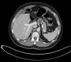
Febrile Lumbar Pain Revealing a Massive Collection: Complicated Psoas Abscess Managed Surgically
Published on: 5th March, 2026
Psoas abscess is a rare but potentially life-threatening condition with non-specific clinical manifestations. The condition may be primary or secondary, depending on whether it has an underlying cause. The condition may be diagnosed with the help of imaging studies, especially contrast-enhanced computed tomography (CT).A 55-year-old female patient with a history of treated hypertension presented with a 15-day history of fever and left-sided low back pain. Her laboratory parameters showed a high leukocyte count of 22,000 cells/mm³ and elevated levels of C-reactive protein (367 mg/L). Her renal functions were within normal limits, and the urine culture was negative. Contrast-enhanced CT revealed a large left-sided intra-abdominal fluid collection extending from the diaphragm to the left iliac fossa, measuring 66 x 305 mm with air bubbles. The collection also showed a left-sided intramuscular psoas collection of 66 x 50 x 131 mm. The patient received intravenous broad-spectrum antibiotics (third-generation cephalosporin, aminoglycoside, and metronidazole). The patient underwent mini-lumbotomy, and nearly 1 liter of pus was drained. The patient’s postoperative course was satisfactory with complete resolution of symptoms. The histopathological examination showed non-specific changes.Psoas abscess should be suspected in patients with febrile low back pain and inflammatory syndrome. Contrast-enhanced CT scans are essential for diagnosis and assessment of the extent of disease. In complicated cases with large abscess formation, early surgical drainage along with appropriate antibiotics will provide the best outcome.This case highlights the importance of early diagnosis and adapted management for successfully navigating the complexities associated with this condition.
Bilateral Severe Encrustation of Long-Term Indwelling Double-J Stents in a Young Non-Lithiasic Patient
Published on: 16th March, 2026
Ureteral double-J stents are a commonly used device in urological practice to allow urinary drainage, avoid ureteral obstruction processes, and protect the upper urinary tract after surgical procedures. However, long indwelling time may give rise to numerous complications, such as infection, migration, and fragmentation of the stent, especially encrustation. Encrustation of stents is a well-known complication that has been closely related to the time active of the stent and can cause significant morbidity if not timely addressed. In severe cases, abundant mineral deposition can result in the development of large calculi encasing the stent and rarely progress to staghorn stones. These cases may pose challenges in terms of the extraction of the stent and may result in complex endourological intervention. The encrustation likelihood is substantially higher if stents are left forgotten or remain in place longer than the advised period. Most patients with heavily encrusted stents have symptoms including flank pain, urinary tract infection, hematuria, or obstructive uropathy, but can present without any symptoms, and this can delay the diagnosis.
Not Every Bladder Mass Is Malignant: A Case of Inverted Urothelial Papilloma in a Young Adult
Published on: 30th March, 2026
Inverted Urothelial Papilloma (IUP) is an unusual variety of urothelial tumors that typically occurs in adults, with a predominance in males. The definitive diagnosis of IUP relies on histopathological examination, as the clinical presentation and endoscopic appearance are non-specific. The recommended treatment for IUP includes the complete transurethral resection, with some controversy regarding the need for cystoscopic follow-up. In this case, we present an atypical instance of IUP in a 21-year-old male patient, diagnosed with gross hematuria and irritative lower urinary tract symptoms. A clinical assessment revealed a 3.0 × 2.6 cm intravesical mass. The definitive diagnosis was confirmed histopathologically and further substantiated by immunohistochemistry, which demonstrated low expression of p53 and Ki-67, effectively ruling out malignancy. This case underscores the diagnostic challenges posed by bladder masses in young adults, emphasizing the necessity of integrating morphological and immunohistochemical findings to prevent overdiagnosis of urothelial carcinoma. The paper focuses on the diagnostic approach and management of this rare condition in the young male population.
Adult Bladder Exstrophy with Premalignant Changes Following Failed Reconstruction: A Case Report
Published on: 1st April, 2026
Background: Bladder exstrophy is a rare congenital abnormality that is usually managed with multiple surgical interventions. Long-term consequences include recurrent urinary tract infections, bladder stones, fistulae, and metaplastic changes with malignant potential.Case Presentation: We present a case of a 21-year-old male with a history of failed childhood surgeries for bladder exstrophy who presented with a vesicocutaneous fistula and a 7 cm bladder stone. He underwent an open cystolithotomy with bladder augmentation and creation of a Benchekroun continent valve. However, the patient developed recurrent fistulae due to poor tissue quality. Histopathological examination confirmed early squamous metaplasia in the bladder mucosa. After discussion in a multidisciplinary meeting, the patient underwent a radical cystectomy with ileal conduit urinary diversion using the Bricker technique. He is doing well at 3 months with no evidence of any complication.Conclusion: This case illustrates the difficulties encountered in managing adult patients with bladder exstrophy and failed reconstructions. The presence of squamous metaplasia, poor bladder tissue, and recurrent complications all contributed to the decision for radical cystectomy. It is important to recognize these changes and address them appropriately in a timely manner to prevent further complications and possible malignant changes.
Burch Colposuspension for Female Stress Urinary Incontinence: A Narrative Review of Contemporary Evidence and Urodynamic Perspectives
Published on: 9th April, 2026
Background: Burch colposuspension is a mesh-free retropubic urethropexy for female stress urinary incontinence (SUI). Amidst increasing scrutiny of synthetic materials, re-evaluating its long-term efficacy and urodynamic profile is essential. Objective: To review contemporary evidence regarding the urodynamic mechanisms, clinical efficacy, and safety profile of the Burch procedure. Methods: A comprehensive literature search was conducted across PubMed, Cochrane Library, and Google Scholar for studies published up to 2025. We included randomized controlled trials, meta-analyses, and long-term cohort studies focusing on Burch colposuspension compared to midurethral slings and autologous slings. Results: Open colposuspension achieves objective cure rates of 68.9%–88% in the first year, with approximately 70% maintaining continence at five years. Long-term studies (mean 13.1 years) show comparable efficacy to midurethral slings (83% vs. 85%). The procedure restores continence by enhancing pressure transmission to the proximal urethra without altering intrinsic sphincter function. While autologous fascial slings offer higher stress-specific success (66% vs. 49%), they carry significantly higher risks of voiding dysfunction requiring reoperation (6.1% vs. 0%). Common complications of Burch include de novo overactive bladder (3%–4.1%) and a higher risk of posterior compartment prolapse (3.3%) compared to slings. Conclusions: Burch colposuspension remains a gold-standard, mesh-free intervention for women with urethral hypermobility, especially those undergoing concurrent abdominal surgery. It provides a durable, safe alternative to synthetic slings with a lower risk of obstructive voiding dysfunction, though patients should be counseled regarding potential long-term pelvic organ prolapse.
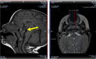
Renal Malakoplakia: A Diagnostic Challenge Presenting as a Subcapsular Collection despite Clinical Recovery
Published on: 9th April, 2026
Background: Renal malakoplakia is a rare chronic granulomatous inflammatory disorder characterized by defective macrophage function. It typically occurs in immunocompromised patients with recurrent urinary tract infections. We present a case of renal malakoplakia in a diabetic patient who progressed to nephrectomy despite initial conservative management.Case presentation: A 57-year-old female patient with a medical history of insulin-dependent type 2 diabetes mellitus was admitted to the hospital with symptoms including fever, left flank pain, and dysuria. A physical examination revealed a tender left lumbar mass. Laboratory investigations revealed a leukocytosis (16,500/mm³), elevated C-reactive protein (142 mg/L), and preserved renal function. A urine culture revealed the presence of multidrug-resistant Escherichia coli (>106CFU/mL). A subsequent Computed Tomography (CT) scan revealed an enlarged left kidney with a 9 × 6 cm multiloculated subcapsular collection, causing significant parenchymal compression, along with two non-obstructive inferior pole calculi. The initial management strategy encompassed ultrasound-guided percutaneous drainage and targeted antibiotic therapy, with the latter being contingent upon bacterial sensitivities. Notwithstanding the patient’s positive clinical recovery, Technetium-99m Dimercaptosuccinic Acid ((99m)Tc-DMSA) renal scintigraphy performed four weeks after the episode revealed a non-functional left kidney, exhibiting a 15% differential function. Following a multidisciplinary discussion, a total left nephrectomy was performed. A histopathological examination revealed extensive replacement of renal parenchyma by polymorphous inflammatory infiltrate with pathognomonic Michaelis-Gutmann bodies. These bodies are spherical, basophilic, perinuclear inclusions that demonstrate strong positivity for Periodic Acid-Schiff and Perls stains. The postoperative course was complicated by self-limited lymphorrhage. At the 3-month follow-up, the patient reported complete resolution of symptoms and remains under nephrological surveillance.Conclusion: This case underscores the diagnostic challenges posed by renal malakoplakia, a condition that can present with a wide spectrum of mimics, including infectious and neoplastic processes. Early diagnosis and prolonged antibiotic therapy with agents capable of intracellular penetration may preserve renal function; however, nephrectomy remains necessary when irreversible parenchymal damage has occurred. Diabetes mellitus has been identified as a significant risk factor for malakoplakia development through impaired leukocyte function.
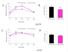
Erectile Dysfunction and Benign Prostatic Hyperplasia: Experience of the Urology Department A of Ibn Sina Hospital: A Prospective Study of 100 Cases
Published on: 24th June, 2026
Background: Erectile dysfunction (ED) and lower urinary tract symptoms (LUTS) due to benign prostatic hyperplasia (BPH) are two common conditions in aging men. Their association exceeds mere age-related coincidence and involves shared vascular, neurological, endocrine-metabolic and psychosexual mechanisms. Objective: To evaluate the prevalence of ED in patients followed for symptomatic BPH, to identify factors associated with its severity, and to analyze the evolution of erectile function after medical treatment. Methods: A prospective cross-sectional study conducted at the Urology Department A of Ibn Sina Hospital, Rabat, from August 1 to November 30, 2021. After excluding 37 records, 100 male patients aged 50 to 80 years followed for BPH were included. LUTS were assessed using the IPSS score and erectile function using the IIEF-5 score. Sociodemographic, clinical, biological, ultrasonographic, cardiovascular and therapeutic data were analyzed. The significance threshold was set at p < 0.05. Results: Mean age was 66 ± 15 years. The overall prevalence of ED was 72%, with 30% mild, 33% moderate, and 27% severe forms among interpretable cases. Only 25% of patients had spontaneously reported their sexual dysfunction. Factors significantly associated with ED were age (p < 0.001), LUTS severity by IPSS (p < 0.001), obesity (p < 0.001), and history of prostatic surgery (p < 0.01), arterial hypertension (p < 0.02) and nocturia (p < 0.05). Alpha-blocker–tadalafil combination therapy improved the IIEF-5 score by +4.3 to +5.2 points. Alpha-blockers alone did not significantly improve erectile function. Conclusion: ED is common and largely under-reported in patients with BPH. LUTS severity, particularly nocturia, is closely linked to erectile impairment. Systematic sexual assessment should be an integral part of BPH management, and the alpha-blocker–PDE5i combination represents a relevant therapeutic strategy in patients with associated ED.
Assessment of the Quality of Life in Patients Undergoing Radical Cystectomy with Urinary Diversion
Published on: 24th June, 2026
Radical cystectomy with urinary diversion is the standard treatment for muscle-invasive bladder cancer. The impact of the type of urinary diversion on quality of life (QoL) remains debated. This cross-sectional study assessed QoL in 57 patients who underwent radical cystectomy between 2014 and 2022 at two urology departments in Rabat, Morocco, using the Bladder Cancer Index (BCI), the SF-36, and the Body Image Scale (BIS). Multiple linear regression identified socioeconomic status, body image perception, urinary discomfort, and comorbidities as the main determinants of QoL, with the type of urinary diversion playing an indirect role mediated through these factors. These findings underscore the need for a multidimensional approach when evaluating and optimizing QoL outcomes after radical cystectomy.
Contact
Select by Volume & Issue
Most Viewed Keywords
- Rothmund-thomson syndrome
- Bilateral sagittal split osteotomy
- Beta-thalassemia major
- Temporomandibular joint
- Nasal cytology
- Depression
- Tooth microchip
- HIV
- WT1
- Venous
- Drug abuse
- Radical cystectomy
- Biological groups of nucleotides
- Clown
- Prevention
- Photonic technology
- Feature processing
- Periodontitis
- Ficus thoningii
- Toxicology
University/Institution
Select and search by University/Institution.
Articles by Country
Select and search by country to get related articles.


Testmonials

I would like to thank JPRA for taking this decision. I understand the effort it represents for you. I'm truly happy to have the paper published in JPRA. And I'll certainly consider JPRA for my next publications as I was satisfied of the service provided, the efficiency and promptness of the interactions we had.
Emmanuel BUSATO
Publishing with the International Journal of Clinical and Experimental Ophthalmology was a rewarding experience as review process was thorough and brisk. Their visibility online is second to none as their published articles appear in all search engines. I will encourage researchers to publish with them.
Elizabeth Awoyesuku
“The choice to submit the forensic case study to the Journal of Addiction Therapy and Research was dictated by the match between the content and the potential readership. The publication process proved to be expedient and we were provided with constructive feedback from reviewers. The final article layout is attractive and conforms to standards. All-in-all, it has been a rewarding process.”
Elisabeth H Wiig
Archives of Vascular Medicine is one of the top class journal for vascular medicine with highly interesting topics. You did a professional and great Job!
Elias Noory
Thank you very much. I think the review process and all of what concerns the administration of the publication concerning our paper has been excellent. The nice and quick answers have been very good I think.
Doris Nilsson
Journal of Pulmonary and Respiratory Research is good journal for respiratory research purposes. It takes 2-3 weeks maximum for review of the manuscript to get published and any corrections to be made in the manuscript. It needs good articles and studies to get publish in the respiratory medicine. I am really glad that this journal editors helped me to get my case report published.
Divya Khanduja
Thanks you and your colleague for the great help for our publication. You always provide prompt responses and high quality of service. I am so happy to have you working with me. Thanks again!
Diana (Ding) Dai
Service and process were excellent as was the “look” of the article when published.
Deane Waldman
Great, thank you! It was very efficient working w/ your group. Very thorough reviews (i.e., plagiarism, peer, etc.). Would certainly recommend that future authors consider working w/ your group.
David W Brett
Your services are very good
Chukwuka Ireju Onyinye
I very much appreciate the humanitarian services provided in my stead by this journal/publisher. It exhibits total absence of editorial impertinence. As an Author, I have been guided to have a fruitful experience. The editorial care is highly commendable.
Chrysanthus Chukwuma
"An amazing experience with the Journal of Advanced Pediatrics and Child Health. Very fast blind review with pertinent corrections and suggestions. I highly recommand both the journal and the editor."
Chaimae Khairoun
The submission is very easy and the time from submission to response from the reviewers is short. Correspondence with the journal is nice and rapid.
Catrin Henriksson
The Clinical Journal of Obstetrics and Gynecology is an open access journal focused on scientific knowledge publication with emphasis laid on the fields of Gynecology and Obstetrics. Their services toward us have been encouraging through their kindness and respect. Great consideration has been given to us as young budding researchers and we are very grateful for this.
Carole Assontsa
During the process your positive communication, prompt feedback and professional approach is very highly appreciated. We would like to thank you very much for your support.
Can Vuran
I do appreciate for your service including submission, analysis, review, editorial and publishing process. I believe these esteemed journal enlighten the science with its high-quality personel.
Bora Uysal
I am very much pleased with the fast track publication by your reputed journal's editorial team. It is really helpful for researchers like me from developing nations. I strongly recommend your journal for publication.
Badri Kumar Gupta
It has been a fabulous journey writing articles for your journal because of the encouragement you people provide for writers from developing nations like India. Kindly continue the same. Looking forward for a long term association.
Badareesh Lakshminarayana
Many thanks for publishing my article in your great journal and the friendly and hassle-free publication process, the constructive peer-review, the regular feedback system, and the Quick response to any queries.
Azab Elsayed Azab
I would like to thank this journal for publishing my Research Article. Something I really appreciate about this journal is, they did not take much time from the day of Submission to the publishing date. Looking forward to have more publications in future.
Ayush Chandra
Submission of paper was smooth, the review process was fast. I had excellent communication and on time response from the editor.
Ayokunle Dada
Your service is very good and fast reply, also your service understand our situation and support us to publication our articles.
Ayman M Abu Mustafa
Really good service with prompt response. Looking forward to having long lasting relationship with your journal
Avishek Bagchi
Your service is excellent. Processing and editing were very fast. I hope to publish more of my works in your journal.
Ausraful Islam
I wanna to thank Clinical Journal of Nursing Care and Practice for its effort to review and publish my manuscript. This is reputable journal. Thank you!
Atsedemariam Andualem
“It was a delightful experience publishing my manuscript with the Clinical Journal of Obstetrics and Gynecology. They offered me lots of opportunities I never had from most publishing houses and their prompt services are greatly appreciated.”
Asafo Jones
I hope to ability to make some new investigation and publish in Your Company in future.
Artur Stopyra
I like the quality of the print & overall service. The paper looks quite impressive. Hope this will attract interested readers. All of you have our best wishes for continued success.
Arshad Khan
Your big support from researchers around the world is the best appreciation from your scientific teams. We believe that there should be no barrier in science and you make it real and this motto come true.
Arefhosseinir Rafi
Your journal co-operation is very appreciable and motivational. I am really thankful to your journal and team members for the motivation and collaboration to publish my work.
Assistant Professor, UCLAS Uttaranchal University, Dehradun, India
Archna Dhasmana
I am glad to submit the article to Heighten Science Publications as it has a very smooth and fast peer-review process, which enables the researchers to communicate their work on time.
Anupam M
This is to specify that I have had an extensive and detailed interaction with the Editorial team of Annals of Clinical Gastroenterology and Hepatology, USA, lasting over a significant period of time. My interaction has been extremely pleasant, especially with Ms Allie Smith, as I find the communication quite inspiring and crystal clear. The attitude of aforesaid individuals is quite helpful and guiding in pertinent instances. It has been a commemorative journey so far working with the Journal and I hope that the symbiosis will continue, evolve and flourish in the forthcoming years. I wish the journal, related personnel and aforementioned individuals a fruitful, successful run.
New Delhi, India
Anubha Bajaj
We appreciate the fact that you decided to give us full waiver for the applicable charges and approve the final version. You did an excellent job preparing the PDF version. Of course we will consider your magazine for our future submissions and we will pay the applicable fees then.
Anna Dionysopoulou
''Co-operation of Archives of Surgery and Clinical Research journal is appreciable. I'm impressed at the promptness of the publishing staff and the professionalism displayed. Thank you very much for your support, help and encouragement.''
Anıl Gokce
Congratulations for the excellence of your journal and high quality of its publications.
Angel MARTIN CASTELLANOS
The service from the journal staff has been excellent.
Andy Smith
I was very pleased with the quick editorial process. We are sure that our paper will have great visibility, among other things due to its open access. We believe in science accessible to all.
Anderson Fernando de Souza
It was a great experience publishing through JCICM. The article has reached out to several institutions. Appreciate your professional work. Hope to work with you again
Anas Wardeh
Publishing an article is a long process, but working with your publication department made things go smoothly, even though the process took exactly 5 months from the time of submitting the article till the time I have favourable response, the missing part is the peer review details, which is essential in self auditing and future improvement, overall experience was excellent giving your understanding of the situation of lack of financial institution support.
Anas Diab
I think that Heighpubs very good. You are very helpful. Thank you for everything.
Ana Ribeiro
Regarding to be services, we note that are work with high standards of professionalism translated into quick response, efficiency which makes communication accessible. Furthermore, I believe to be much inviting for the submission of future works for publication purposes.
Amélia João Alice Nkutxi
I would like to mention that I had a wonderful experience working with HSPI. The whole process right from manuscript submission to peer review till the publication of the article was very prompt & efficient. I wish you good luck for the future.
Amarjeet Gambhir
Once I submitted the manuscript, the response time of the reviewers was very fast. The fine-tuning of the galley proof was likewise prompt. I believe the journal provide a valuable outlet to disseminate physical rehabilitation scientific knowledge to the clinical community. Respectfully. Dr. Alon
Alon
We really appreciate and thanks the full waiver you provide for our article. We happy to publish our paper in your journal. Thank you very much for your good support and services.
Ali Abusafia
It was a real pleasure working with your team. The review was done fast, and it was very clear, the editing was flawless, the article was published quickly compared to other journals, and everyone was understanding and helpful. I will gladly recommend this journal to my acquaintances in academia.
Alexandra Cozma
To the editorial team at HSPI and the Journal of Clinical Nephrology: Thank you so much for your hard work and collaboration in bringing our article to life. Your staff was responsive, flexible, and communicative and made the process smooth and easy. Thank you!
Alejandro Munoz
Dear colleagues! I am satisfied with our cooperation with you. Your service is at a high level. I hope for a future relationship. Let me know if I can get a paper version of the magazine with my articles from you. I see them on the Internet.
Aksenov V.V
"This is my first time publishing with the journal/publisher. I am impressed at the promptness of the publishing staff and the professionalism displayed. Thank you for encouraging young researchers like me!"
Ajite Kayode
I want to thank you for our collaboration. You were fast and effective with a positive spirit of teamwork. I am truly excited from our collaboration. You were like always fast, efficient and accurate. I hope that in the near future we will collaborate again.
Aikaterini Solomou
In my opinion, you provide a very fast and practical service.
Ahmet Eroglu
Great, We are too comfortable with the process including the peer review process and quality. But, the journal should be indexed in different databases such scopus.
Afework Edmealem
We really appreciate your efforts towards our article, the professional way you handle our request for exemption from charges. It was a great honor for us to publish in your magazine.
Achraf elbakkaly
I really liked the ease of submitting my manuscript in the HSPI journal. Further, the peer review was timely completed and I was communicated the final decision on my manuscript within 10 days of submission which is really appreciable. I strongly recommend all the scientists and researchers to submit their work in this journal”
Abu Bashar
My candid opinion is that the service you render is second to none. My favourite part is the prompt response to issue, really i value that.
Abiodun Akanbi Adeogun
Thank you very much for accepting our manuscript in your journal “International Journal of Clinical Virology”. We are very thankful to the esteemed team for timely response and quick review process. The editorial team of International Journal of Clinical Virology is too cooperative and well-mannered during the publication process. We are hopeful to publish many quality papers in your journal and I suggest the International Journal of Clinical Virology to all of my colleagues, researchers and friends to publish their research here.
Abdul Baset
I, Muhammad Sarwar Khan, am serving as Editor on Archives of Biotechnology and Biomedicine (ABB). I submitted an editorial titled, 'Edible vaccines to combat Infectious Bursal Disease of poultry' for publication in ABB. After submitting the manuscript; the services rendered by the management and technical personnel to handle and process the manuscript were marvelous. Plagiarism report was shared with me with complements before reviewers' comments, All steps including article processing and service charges were well taken care of keeping in view the author's interest/preference. All together, it was an encouraging and wonderful experience working with ABB personnel.
University of Agriculture, Pakistan
Muhammad Sarwar Khan
Your journal has accomplished its intended mission of providing very effective and efficient goals in dealing with submissions, conducting the reviewing process and in publishing accepted manuscripts in a timely manner. Keep up the great work and services that you provide.
University of Jacqmar, Inc., USA
John St. Cyr
I am to express my view that Heighten Science Publications are reliable quick even after peer review process. I hope and wish the publications will go a long way in disseminating science to many interested in scientific research.
College of Fisheries, CAU(I), Tripura, India
Ajit Kumar Roy
The Journal Clinical Nephrology provides a good opportunity for readers to stay updated in the field of clinical nephrology. Additionally - it provides a good opportunity for authors to publish their work. 1. Publication of the accepted manuscripts is sufficiently rapid. 2. The trust factor between the journal and me, as an author, is very important and well preserved. 3. Peer review process very rapid and effective.
Assaf Harofeh Medical Center, Israel
Leonid Feldman
In 2017, I submitted a manuscript to the journal Archives of Biotechnology and Biomedicine belonging to Heighten Science Publications Corporation. Within one week I already received the response from the editor. All processing steps were really fast so in terms of a speedy publication I can particularly recommend the journal Archives of Biotechnology and Biomedicine. The responsible contact person of the journal was always available, which gives a trustworthy impression to the author. Also the peer review process was clear and constructive. So from my experience with Heighten Science Publications Corporation I can recommend publishing there.
University of Tubingen, Germany
Yvonne Mast
We thank to the heighten science family, who speed up the publication of our article and provide every support.
Mehmet Besir
The services of the journal were excellent. The most important thing for an author is the speed of the peer review which was really fast here. They returned in a few days and immediately replied all of my questions. I want to refer this platform to all scholars. Many thanks.
Eastern Mediterranean University, Cyprus
Zehra Guchan TOPCU
Thank you for your attitude and support. I am sincerely grateful to you and the entire staff of the magazine for the high professionalism and fast quality work. Thank you very much!
USA
Igor Klepikov
Thank you and your company for effective support of authors which are very much dependable on the funds gambling for science in the different countries of our huge and unpredictable world. We are doing our work and should rely on a teams like Galley Proof-HSPC. Great success to all of you for the 2019th! Be well all the year long.
Russia
Victor V Apollonov
The editorial process was quickly done. The galley proof was sent within a week after being accepted for publication. The editorial team was very helpful and responded promptly.
India
Rohit Kulshrestha
Publishing with the International Journal of Clinical and Experimental Ophthalmology was a rewarding experience as review process was thorough and brisk. Their visibility online is second to none as their published articles appear in all search engines. I will encourage researchers to publish with them.
University of Port Harcourt Teaching Hospital, Nigeria
Dr. Elizabeth A Awoyesuku
"It was a pleasure to work with the editorial team of the journal on the submission of the manuscript. The team was professional, fast, and to the point".
NC A&T State University, USA
Moran Sciamama-Saghiv
Submission of paper was smooth, the review process was fast. I had excellent communication and on time response from the editor.
Ekiti State University Teaching Hospital, Nigeria
Ayokunle Dada
I am delighted and satisfied with. Heighten Science Publications as my manuscript was thoroughly assessed and published on time without delay. Keep up the good work.
Ido-Ekiti/Afe Babalola University, Nigeria
Dr. Shuaib Kayode Aremu
"This is my first time publishing with the journal/publisher. I am impressed at the promptness of the publishing staff and the professionalism displayed. Thank you for encouraging young researchers like me!"
Ekiti State University, Nigeria
Adebukola Ajite
I wanna to thank clinical journal of nursing care and practice for its effort to review and publish my manuscript. This is reputable journal. Thank you!
Wollo University, Ethiopia
Atsedemariam Andualem
We appreciate your approach to scholars and will encourage you to collaborate with your organization, which includes interesting and different medical journals. With the best wishes of success, creativity and joy in life, prosperity in the medical field.
Ivano- Frankivsk National Medical University, Ukraine
Nataliya Kitsera
Thank you very much for your support and encouragement. I am truly impressed by your tolerance and support. Thank you very much
Diaverum: PADC, Jeddah, Saudi Arabia
Nasrulla Abutaleb
You are such a nice person. Your journal co-operation is very appreciable and motivational.
Department of Biotechnology, Uttaranchal college of Applied and Life Sciences, Uttaranchal University, Dehradun, Uttarakhand, India
Archna Dhasmana
“Mobile apps and wearable technology are becoming ubiquitous in our environment. Their integration with healthcare delivery is just beginning to take shape. The early results are promising and the possibilities great."
BS, PharmD., MBA, CPHIMS, FHIMSS, Adjunct Professor, Global Healthcare Management, MCPHS University, Chief Strategy Offi cer, MedicaSoft, Senior Advisor, National Health IT (NHIT) Collaborative for Underserved, New York HIMSS, National Liaison, Health 2.0 Boston, Past Chair, Chair Innovation, USA
Helen Figge
“The choice to submit the forensic case study to the Journal of Addiction Therapy and Research was dictated by the match between the content and the potential readership. The publication process proved to be expedient and we were provided with constructive feedback from reviewers. The final article layout is attractive and conforms to standards. All-in-all, it has been a rewarding process.”
Ph.D, Boston University Department of Communication Sciences and Disorders and Knowledge Research Institute, Inc., 2131 Reflection Bay Drive, Arlington, Texas 76013, USA
Elisabeth H. Wiig
The service is nice and the time of processing the application is fast.
Department of Neurosurgery, Queen Elizabeth Hospital, Hong Kong
Long Ching
Your service is very good and fast reply, Also your service understand our situation and support us to publication our articles.
Palestine College of Nursing, Khan Younis, Gaza Strip, Palestine
Ayman M Abu Mustafa
“It was a delightful experience publishing my manuscript with the Clinical Journal of Obstetrics and Gynecology. They offered me lots of opportunities I never had from most publishing houses and their prompt services are greatly appreciated.”
Department of Agricultural Economics, Agribusiness and Extension, Kwame Nkrumah University of Science and Technology, Kumasi, Ghana
Akowuah Jones Asafo
Related Journals

If you are already a member of our network and need to keep track of any developments regarding a question you have already submitted, click "take me to my Query."